Male Reproductive Histology Review - Practical 3

1. Identify the following structures in the photo below.
A.
Ductus deferens 
Testis tissue 
Epididymis  
Prostate  
B.
Ductus deferens 
Testis tissue 
Epididymis 
Prostate 
C.
Testis 
Epididymis 
Seminiferous tubule 
Interstitial Cells 
D.
Cells of Leydig 
Seminiferous tubule 
Testes tissue 
Urethra 
E.
Urethra 
Testes tissue 
Seminiferous tubule 
Cells of Leydig 


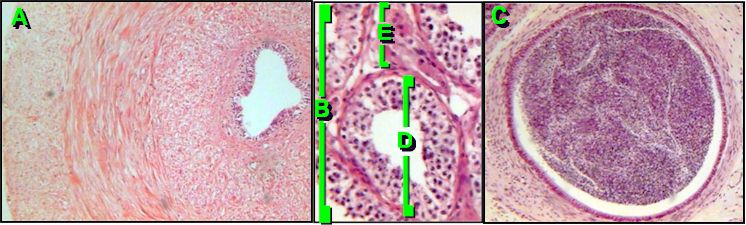
2. Identify structues in the photo below.

A.
Kidney 
Penis
Liver 
Bladder 
B.
Corpus cavernosum 
Prepuce 
Corpus spongiosum 
Urethra 
C.
Corpus cavernosum 
Prepuce 
Corpus spongiosum 
Urethra 
D.
Corpus cavernosum  
Prepuce  
Corpus spongiosum  
Urethra  


3. Name the parts of the photo below.
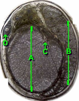
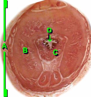
A.
Epididymis 
Ductus deferens  
Testis 
Prostate  
B.
Epididymis 
Ductus deferens 
Testis 
Rete testis 
C.
Epididymis 
Ductus deferens 
Testis 
Rete testis 
D.
Epididymis 
Ductus deferens 
Testis 
Rete testis 


AP II Lab | APLabs Homepage