Large Intestine Microscopic Cross Section
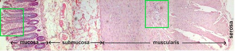
 Mucosal layer on the surface is made up simple columnar cells and a mucosal muscularis on the deep side . Mucosal layer on the surface is made up simple columnar cells and a mucosal muscularis on the deep side . |
| Submucosa contains fibrous connective tissue and blood vessels. |
| The muscularis externa is made up of a circular and a longitudinal muscle layer with a myenteric plexus in between the layers. |
| A very thin layer of Serosa is also present . |
| Click on the green squares to see details of the muscosa and muscularis externa. |